
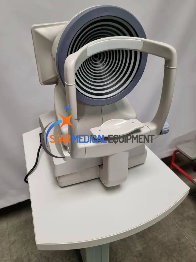
Topcon CA-800 Corneal Topography
Used Topcon CA-800 Corneal Topography Contact Lens and Dry Eye Analyzer
- Used Condition
- Genuine Product
- Manufacturer Warranty
- Technical Support
- Worldwide Shipping
Selling a used 2017 Topcon CA-800 Corneal Topography and Dry Eye Workstation is in excellent condition. Comes with a 30-day money back guarantee. We take responsibility until the packages reach destination with protected, robust wooden crating system and ships worldwide.
The CA-800 allows for complete evaluation of the anterior surface and tear related structures.
Key Features
- Topography Map, Corneal Wavefront (Zernike) Analysis
- Tear Film Breakup Tear Meniscus Height/Blink Analysis
- Comparison Map, Differential Map, Corneal Height Map
- Contact Lens Fitting Simulation
- White to White Measurement
- Pupillometry
- Keratoconus Screening
- Meibomian Gland Analysis
- PC Integrated, Space Saving
ALL FEATURES ACCESSIBLE ON JUST ONE SCREEN
Acquisition
The CA-800 is easy to use. Visual signals support fast and easy alignment and accurate focusing.
The CA-800 has Right and Left eye detection, preventing incorrect filing of data to the wrong eye.
The automated best image selection mode in the CA-800 software, selects the best focused position and automatically acquires the image.
Corneal comparison & differential map
With the CA-800, it is easy to compare topography maps between two examinations of the same patient to examine changes over time. Parameters such as keratometry, apical curvature and corneal symmetry can be analyzed to monitor any corneal surface changes, for example caused by keratoconus. With the differential map, the effect of a procedure such as refractive surgery, can be observed. It is also useful to examine the response of the cornea toan orthokeratology lens.
Corneal Zernike analysis
The Zernike analysis module provides 36 polynomials up to the 7th order, giving a clear view of the optical aberrations which can disturb vision. Based on this information, the CA-800
simulates the effect on vision, which is useful for educating patients about their condition.
The pupil size can be selected to examine the implications of the corneal aberrations under different light levels.
Contact lens fitting simulation
The CA-800 provides a comprehensive platform to optimize contact lens fitting. On-board simulation software automatically selects the best fitting contact lens based on an internal contact lens database for all the main manufacturers (upgradable and customizable by the user). Lens parameters and position can be modified, and the resulting fluorescein simulation
saved to a library, to allow rapid comparison between lens options. This minimizes the number of lenses that need to be trialed on the patient’s eye, saving time and making the process more acceptable for the patient.
Fluorometry
The CA-800 incorporates eight blue LED’s for fluorescein imaging and real time fluorescein movies which are valuable for contact lens fitting and dry eye assessment. During every measurement, the CA-800 automatically registers the pupil diameter, for comparison with the size and location of the contact lens optical zone. Real time fluorescein movies allow the eye
care practitioner to judge the movement of the contact lens on the cornea, the distribution of the tear film under the contact lens as well as the wetting of the anterior surface of the contact
lens. Staining of the ocular surface can also be recorded as images or a movie. The tear film condition, corneal artifacts and fluorescein break up time (BUT) can be observed.
Pupillometry
The CA-800 is equipped with different LED lights to support dynamic and static pupillometry under a range of lighting conditions. With the CA-800, the user can check the pupil position and diameter (under photopic through to scotopic lighting conditions) in relation to the position of the optical zone of an Ortho-K treatment, contact lens, intraocular lens or laser treatment. Dynamic pupillometry provides clear information on the reaction time of the pupil along with changes in pupil center location.
Keratoconus screening
With the CA-800, signs of corneal asymmetry of the cornea can easily be detected even at an early stage. By analyzing various characteristics of the corneal shape, a keratoconus probability index is calculated, with color coding to indicate the level of compatibility of each metric with keratoconus.
The CLMI (Cone Location and Magnitude Index) is available as an alternative. These metrics along with comparison maps, make it easy to monitor keratoconus and keratoconus-like patterns over time.
Meibomian gland analysis
With the Infra-red illumination of the CA800, the Meibomian glands of the upper and lower eyelid can be captured and analyzed.
Posterior blepharitis is the most common form of lid margin disease. MGD (Meibomian Gland Dysfunction) can cause or exacerbate dry eye symptoms and eyelid inflammation.
The oil glands become blocked with thickened secretions. Chronically clogged glands eventually become unable to secrete oil which results in permanent changes in the tear film resulting
in ocular surface disease. With the CA-800, Meibomian glands can easily be observed, to inform management decisions and encourage compliance with treatment.
DRY EYE SUITE
Blink analysis
The blink analysis function records normal blinking over a period of time to automatically calculate the average blinks per minute and inter-blink interval.
Combining this data with the non-invasive tear break up measurement allows the Ocular Protection Index (OPI) to be calculated, identifying eyes at risk of ocular surface damage.
Non-invasive breakup time
The non-invasive breakup time (NIBUT) records the patient’s tear film condition while they hold their blink. It is the Gold Standard for assessing tear breakup, as recommended by DEWS II*. The corneal surface is divided into sectors with breakup defined as the time for 5% of all sectors to demonstrate breakup. Repeat measurements can be averaged, also allowing areas of recurrent first break to be identified. In addition to these informative metrics, the video playback feature allows visualization of the tear breakup over time, along
with associated changes in topography and anterior surface aberrations.
Tear meniscus height
Detailed images of the inferior tear meniscus can be captured with the CA-800. During review, touching the meniscus adds a marker based on automatic detection of the lid margin and the top of the tear meniscus. The position, separation and angle of each marker can be fine tuned if required. The tear meniscus profile is then displayed along with relevant metrics: minimum, maximum, average and variation.
ADDITIONAL FEATURES
i-Map
The optional software i-Map enables delegation of acquisition to support staff while the clinician reviews the data in another room. i-Map allows full manipulation of the data. It contributes
to optimizing your clinical workflow.
DICOM™ Compliance
The DICOM panel in the CA-800 connectivity settings allows the user to set the required parameters for the connections to the available DICOM features:
- Modality Worklist
- Patient Root Query
- Storage
- Storage Commitment
Zeiss Axio Observer 3 Specifications
| Keratoscope cone | 24 rings equally distributed on a 43D sphere |
| Analyzed points | Over 100.000 |
| Measured points | 6.200 |
| Corneal coverage | Up to 9.8mm on a sphere of radius 8.00mm (42.2 diopters with n=1.3375) |
| Diopter power range | From 1D to 120D |
| Resolution | +/- 0.01D, 1 micron |
| Accuracy / Precision axial radius | +/- 0.03mm altimetric data, +/- 2µm at 4mm |
| Capture system | Guided focus with auto-capture |
| Output ports | 2xUSB, LAN |
| Monitor | LCD 10.1 inch capacitive touch screen |
| Database | Internal |
| Pupillometry | Dynamic, Photopic, Mesopic, Scotopic |
| Fluorescein | Image, Video |
| Report | Corneal map, Comparison map, Contact lens, Height map, Zernike analysis, Pupillometry, Toric IOL, Screenshot, NIBUT, Meibomian gland analysis, TMH, Fluorescein |
| Working environment | 10°-40°C, Relative humidity 30-75% (no dewing), Atmospheric pressure 700-1060hPa |
| Power source | AC 100-240V 50/60 Hz |
| Power consumption | 80 VA |
| Connections | Wi-Fi Optional, LAN integrated / iMAP |
| Printing options | USB printer, Network printer, PDF on network shared folder, PDF on USB |
| Operating System | Windows 10 Enterprise LTSC (Long Term Support Channel) |
| RAM | 4GB |
| Hard Disk | At least 500GB SATA Hard drive (database storage) 32 GB Solid State Drive (operating system and application software) |
| Dimensions (W x H x L) | 320 x 490 x 470 mm |
| Weight | 15 Kg |