
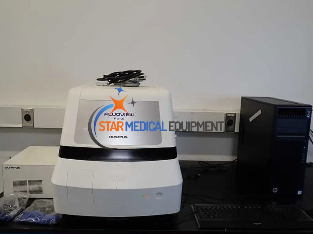
Olympus FLUOVIEW FV10i Confical Laser Scanning
Olympus FLUOVIEW FV10i Confical Laser Scanning Biological Microscope System
- New Condition
- Genuine Product
- Manufacturer Warranty
- Technical Support
- Worldwide Shipping
Up for sale a used Olympus FLUOVIEW FV10i Confical Laser Scanning FV10C-O with FV10C-PSU and HP Z440 Computer (Olympus FV10-ASW Software installed Version 04.02). Item is in great technical. Comes with a 30-day money back guarantee. We take responsibility until the packages reach destination with protected, robust wooden crating system and shipped worldwide.
Package includes:
- Olympus FV10i unit
- FV10C-PSU (100-240 V, 50/60 Hz)
- Three Glass bottom dishes (33mm diameter) plate
- Well slide (8 wells type) plate
- Culture pod (35mm diameter) plate
- HP Z440 Computer with Monitor, Mouse and Keyboard
The unique FLUOVIEW FV10i-LIV is a fully automated confocal laser-scanning microscope. The completely re-engineered design of this microscope, integrated into a self-contained package with a variety of functions, enables even inexperienced and first-time users to perform easily and efficient high-quality confocal imaging. Olympus has made no compromises in ergonomics and image quality, using high-quality optical components and smart and easy software.
Olympus FV10i-LIV Specifications
| Laser Light Source | |||||||||||
| LD lasers | 405nm(17.1mW),473nm(11.9mW),559nm(15mW),635nm(9.5mW) | ||||||||||
| Modulation | Continuously Variable by the LD direct modulation (0.1%-100%, 0.1% inclement) Line return period - laser OFF | ||||||||||
| Scanning | |||||||||||
| Scanning method | 2 galvanometer scanning mirrors | ||||||||||
| Scanning mode |
|
||||||||||
| Detection | |||||||||||
| Detector module | Fluorescence: 2 channels, Phase Contrast: 1 channel | ||||||||||
| Variable barrier fi lter mechanism for fl uorescence channel by diffraction grating and slit | |||||||||||
| Detection method | Analog integration detection by Photomultiplier | ||||||||||
| Pinhole | Single motorized pinhone | ||||||||||
| Pinhole diameter: ø50-800µm automatic setting (adjustable to x1.0, x1.5, x2.0, and x2.5) | |||||||||||
| Field number | 18 | ||||||||||
| Optical zoom | 10x objectives: 1x – 6x in 0.1x increments | ||||||||||
| 60x objectives: 1x – 10x in 0.1x increments | |||||||||||
| Automatic Exposure | Automatic setting of the laser intensity and photomultiplier sensitivity to fl uorescence intensity | ||||||||||
| Focus | |||||||||||
| Z-drive | Motorized focus | ||||||||||
| Minimum increment: 0.01µm | |||||||||||
| Objectives | Exclusively designed 10x phase contrast objective / NA 0.4 (equivalent to UPLSAPO 10x) | ||||||||||
| Exclusively designed 60x phase contrast water-immersion objective / NA 1.2 (equivalent to UPLSAPO 60x W) / with motorized correction collar | |||||||||||
| Remote switching from software by electric revolver | |||||||||||
| Automatic focus (AF) | Automatic detection of interface between specimen and cover glass by laser refl ection light detection | ||||||||||
| Automatic detection of cover glass thickness and automatic setting of motorized correction collar | |||||||||||
| Water supply | Automatic water supply and air cleaning mechanism for 60× Water-immersion objective | ||||||||||
| XY Stage | |||||||||||
| XY driving method | Motorized XY stage module by stepping motor | ||||||||||
| Minimum increment: 0.3µm | |||||||||||
| Specimen holder | Only the dedicated specimen holder can be mounted | ||||||||||
| For three glass bottom dishes with 35mm diameter | |||||||||||
| For a glass slide, For one set of cover glass chamber (8 wells type) | |||||||||||
| For Well slide (8 wells type),Culture pod(for a glass bottom dish with 35mm diameter) | |||||||||||
| Incubator | |||||||||||
| Room environmen |
|
||||||||||
| Heating method | Non-contact heating by resistive heater mounted on frame section | ||||||||||
| Control Device | |||||||||||
| Controller | Dedicated controller PC/AT-compatible | ||||||||||
| OS: Windows Vista Business, 32 bit (English version), CPU: Intel Core2Duo 3.0GHz | |||||||||||
| RAM: 2GB × 2, HDD: 500GB × 2, Special PCI-Express I/F board built-in, Optical drive: DVD-Multi drive built-in | |||||||||||
| Main Software Feature | |||||||||||
| Image acquisition mode | Map image, one shot, time-lapse (XYT), Z-stack (XYZ), Z-stack time-lapse (XYZT), multi area time-lapse (Multi Area XYT), multi area Z-stack time-lapse (Multi Area XYZT) | ||||||||||
| Specimen setting | Automatic setting for fl uorescence channel and laser according to Dye selected from Dye list | ||||||||||
| Map image acquisition | Automatic selection of map image of 3×3 – 9x9 fi elds according to 10× objective lens (The maximum area varies in accordance to the specimen holder used), and manual selection of map acquisition area | ||||||||||
| Multi area time-lapse | Automatic multi area time-lapse by motorized XY stage | ||||||||||
| Setting for each registered point: Image size, scanning speed, cross talk reduction, pinhole diameter, rotation angle, galvano zoom, acquisition channel, laser power, PMT sensitivity, Z condition | |||||||||||
| Maximum resister number: 10 items per one container | |||||||||||
| Maximum interval time: one hour | |||||||||||
| Maximum acquisition number of times: 3000 times per one point | |||||||||||
| Image acquisition area | Area appointment: All area, clipping square area (minimum area: 96 × 96 pixels) | ||||||||||
| Image display | Display by channel, overlapping display, image in progress review | ||||||||||
| Cross talk reduction | Line sequential action (2 channel), or frame sequential action (3 channel and 4 channel) | ||||||||||
| Acquisition image fi le type | OLYMPUS image format (OIF) | ||||||||||
| Image file type available for viewing | OLYMPUS image format (OIF, OIB), Multi-TIFF format (8/16 bit grey scale, index color, 24/32/48 bit color), JPEG, BMP, TIFF | ||||||||||
| Image editing | LUT: pseudo color setting, contrast adjustment, Comment: inputting graphic, text, scale etc., image extraction, combination | ||||||||||
| 3D image construction | 3D display: AlphaBrend method, Maximum intensity projection method 3D animation display, free orientation of cross section display | ||||||||||
| Image processing | Various types of image fi lter: Median, Enhanced Edge, etc. | ||||||||||
| Calculations: inter-image, arithmetic and logical operation | |||||||||||
| Image analysis | Area and perimeter measurement, time-lapse measurement, colocalization analysis | ||||||||||
| Room Environment | |||||||||||
| Temperature | 18-28˚C(fl uctuation ±2˚C) | ||||||||||
| Humidity | 30-80% (non condensing) | ||||||||||