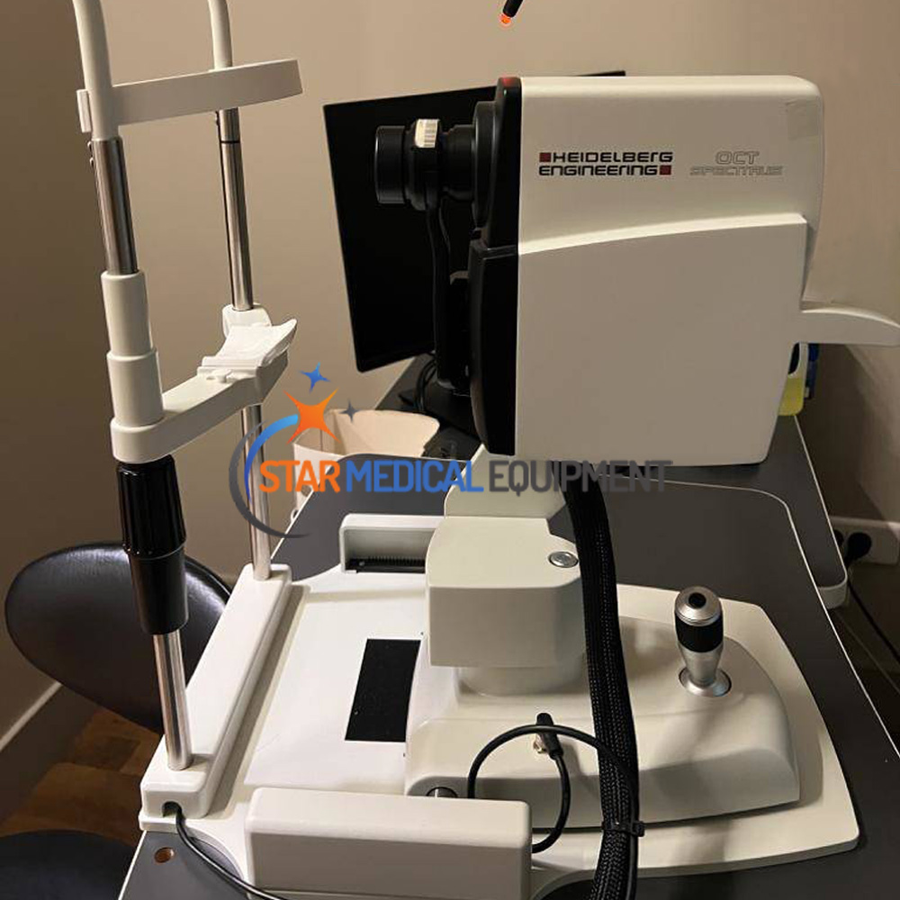
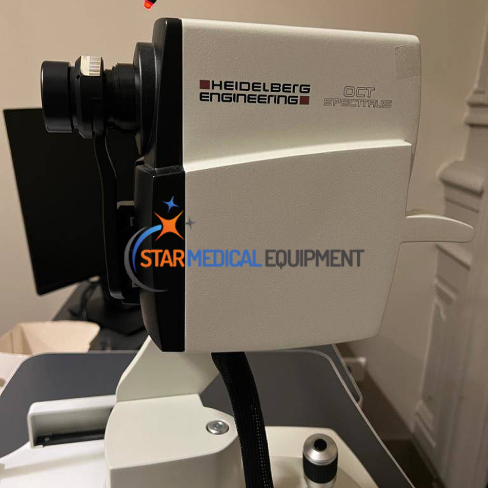
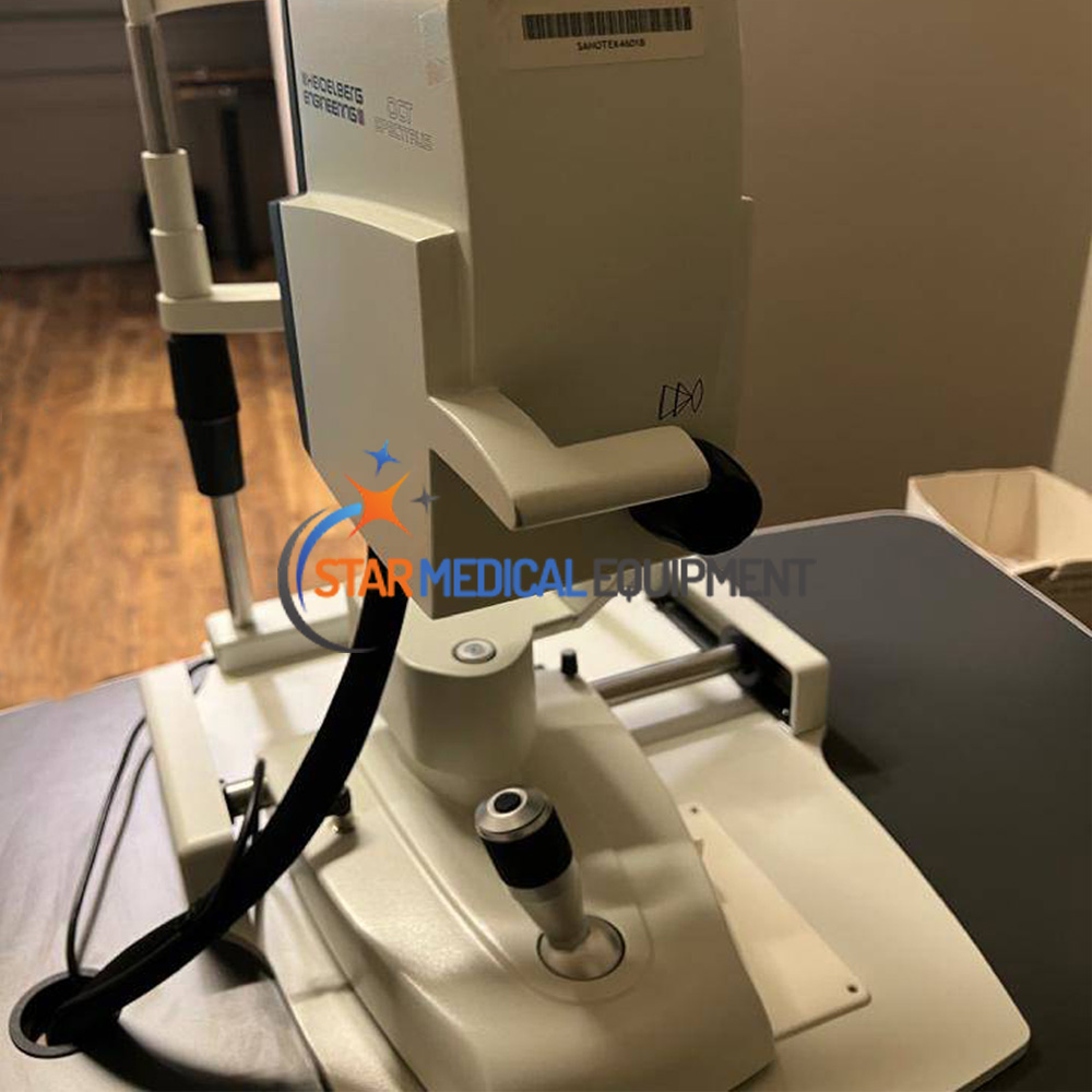
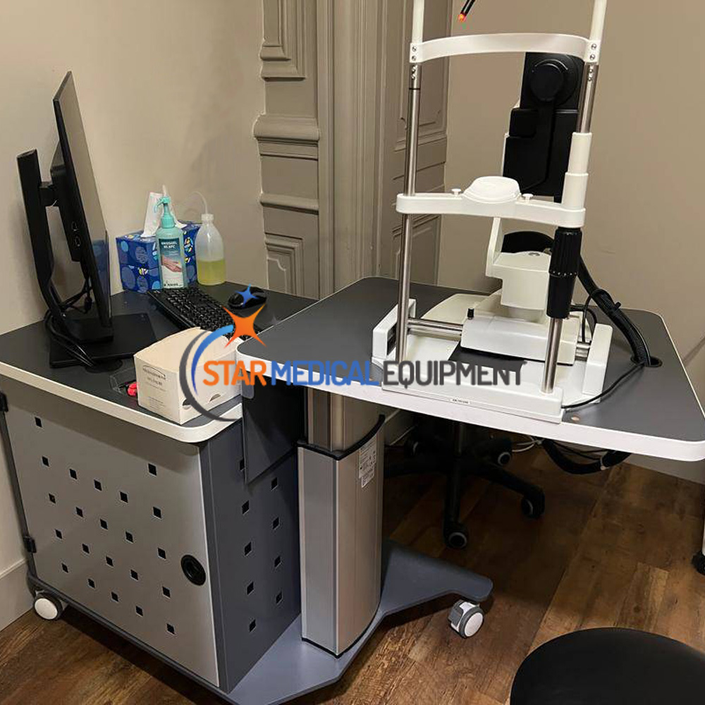
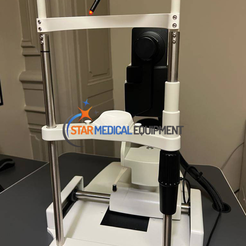
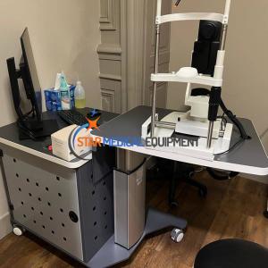
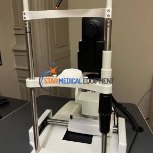
Heidelberg Spectralis OCT2
Sale Heidelberg Spectralis OCT2 (OCTA) Retinal Imaging System. Used in great condition.
$27,000
Up for sale used Heidelberg Spectralis OCT2 is in great condition, it is in As new condition. Comes with a 6 months warranty and 30 days money back gaurantee.
It offers great performances for Retina and Glaucoma, combining scanning laser fundus imaging and simultaneous High Resolution OCT.
It uses the patented technologies : TruTrack Active Eye tracking technology, Heidelberg noise reduction, AutoRescan, Confocal scanner Laser Ophtalmoscopy. It also proposes an Anatomic positioning system.
Imaging capabilities: OCTA (Angiography without injectrion) Fundus Camera, Wide-Field Imaging, Retina Thickness, Retinal Mapping, RPE Deformation.
Powerful diagnostic imaging configuration combines high speed SD-OCT with 5 fundus imaging modes for maximum detection, diagnosis and disease management information. Upgradable platform with panning camera head
The SPECTRALIS® OCT2 is an ophthalmic imaging platform with an upgradeable and modular design. This flexible platform allows clinicians to configure each SPECTRALIS to the specific diagnostic workflow in the practice or clinic.
Core Technologies
The SPECTRALIS OCT2 platform is based on 5 core technologies:
cSLO
The SPECTRALIS OCT uses confocal scanning laser ophthalmoscopy (cSLO) for fundus and anterior segment imaging. The confocal principle minimizes the effects of scattered light to produce high-contrast and detailed images. In many cases, a comprehensive assessment of the retina is possible even in patients with cataracts.
SD-OCT
Spectral domain optical coherence tomography (SD-OCT) provides high-resolution, two-dimensional OCT images of the retina and anterior segment. The next generation SD-OCT technology (OCT2) offers enhanced image quality from vitreous to choroid and faster image acquisition with a scanning speed of 85 kHz for improved clinical workflow.
TruTrack Active Eye Tracking
TruTrack Active Eye Tracking is a patented technology that utilizes two laser scanning systems simultaneously to actively track the eye in real time throughout image acquisition. This mitigates the effects of eye motion, resulting in high-resolution OCT images. TruTrack is indispensable for the acquisition of high quality images throughout a volume scans.
Noise Reduction
TruTrack Active Eye Tracking enables the capture of multiple OCT images in the exact same location. With these multiple images, SPECTRALIS noise reduction technology is able to differentiate structural information from noise, reducing noise even further and resulting in high contract images.
AutoRescan
With the AutoRescan function, follow-up examinations are automatically and precisely aligned with the fundus image of the reference examination using anatomic landmarks that allow for the detection of even very small pathologic changes over time. Studies have shown that the AutoRescan function offers reliable retinal thickness measurements as small as 1 micron.
Multimodal Imaging
SPECTRALIS facilitates comprehensive diagnostics by combining multiple acquisition modes in a single device.
Infrared-Reflectance (IR)
With a wavelength of 815 nm, infrared reflectance allows for comfortable imaging and exact, high-resolution visualization of intraretinal changes, such as cystoid macular edema (CME) or central serous chorioretinopathy (CSCR).
Blue-Reflectance (red-free)
A blue laser with a wavelength of 486 nm is used for the acquisition of red-free fundus images. Blue reflectance images are best for spotting pathological changes in superficial retinal structures, such as epiretinal membranes, retinal folds, and cysts.
MultiColor
MultiColor simultaneously uses three laser wavelengths (infrared, green, and blue) to provide diagnostic images that show distinct structures at different depths within the retina. These detailed images enable visualization of structures and pathology not always visible on ophthalmoscopy or traditional fundus photography.
Scanning Laser Angiography
SPECTRALIS scanning laser angiography can be conducted with either fluorescein or ICG dye. Detailed, high-resolution images and video sequences visualize vessel filling, flow, and leakage. FA and ICGA can be acquired simultaneously or individually in combination with OCT imaging.
Fundus Autofluorescence (BluePeak)
BluePeak reveals metabolic stress in the retina using fluorescent lipofuscin as an indicator. With non-invasive fundus autofluorescence, areas of geographic atrophy and other pathologies can be easily visualized. The optional RegionFinder software allows for the quantification of atrophic areas in the retina seen on BluePeak images.
OCT Angiography (OCTA)
The OCT Angiography Module is a non-invasive imaging method that produces detailed three-dimensional representations of the retinal and choroidal vasculature. The multi-modal SPECTRALIS platform offers the ability to combine OCTA with structural OCT, confocal scanning laser imaging, and dye-based angiography in a single device.
Spectralis OCT2 Technical Data
| General Information | |
| Input voltage | 100 – 240 Volt |
| Power consumption | 140 VA |
| Light Sources | |
| FA laser | 486 nm (blue) |
| Green laser | 518 nm (green) |
| ICGA laser | 786 nm (infrared) |
| R laser | 815 nm (infrared) |
| OCT-SLD | 880 nm (infrared) |
| Standard Objective Lenses | |
| Max. scan field (visual angle) | 30° × 30° (ca. 9 × 9 mm) |
| ART (image averaging) | up to 100 single images |
| Widefield Imaging Objective Lens | |
| Max. scan field (visual angle) | Ø 55° (ca. 16,5 mm) |
| High resolution mode (digital) | ≈ 10 μm/pixel lateral |
| ART (image averaging) | up to 100 single images |
| Ultra-Widefield Imaging Objective Lens | |
| Max. scan field | Ø 102° (ref. pupil), Ø 135° (ref. eye center) |
| High resolution mode (digital) | ≈ 20 μm/pixel lateral |
| ART (image averaging) | up to 100 single images |
| OCT – Retinal imaging | |
| A-scan rate | 85 kHz |
| Axial digital image size | 496 pixel |
| Axial resolution | 3,9 μm/pixel |
| Lateral resolution | 5,7 μm/pixel |
| Scan angle | up to 55° (ca. 16,5 mm) |
| Scan depth into the tissue | 1,9 mm |
| Scan patterns | Line scan, volume scan, radial scan, circle scan, ONH-RC scan, individual |
| OCT – Anterior imaging | |
| cSLO image width | 8,3 / 11 / 16,6 mm |
| OCT scan depth | 8,3 / 11 / 16,6 mm |
| ART (image averaging) | up to 100 single images |
| OCT Angiography imaging | |
| High speed mode | 30° × 15° with 11 μm lateral resolution |
| High resolution mode | 10° × 10° with 5,7 μm lateral resolution |
| Number of pixel | Max. 768 × 384 pixel |
| ART (image averaging) | up to 7 single images |
