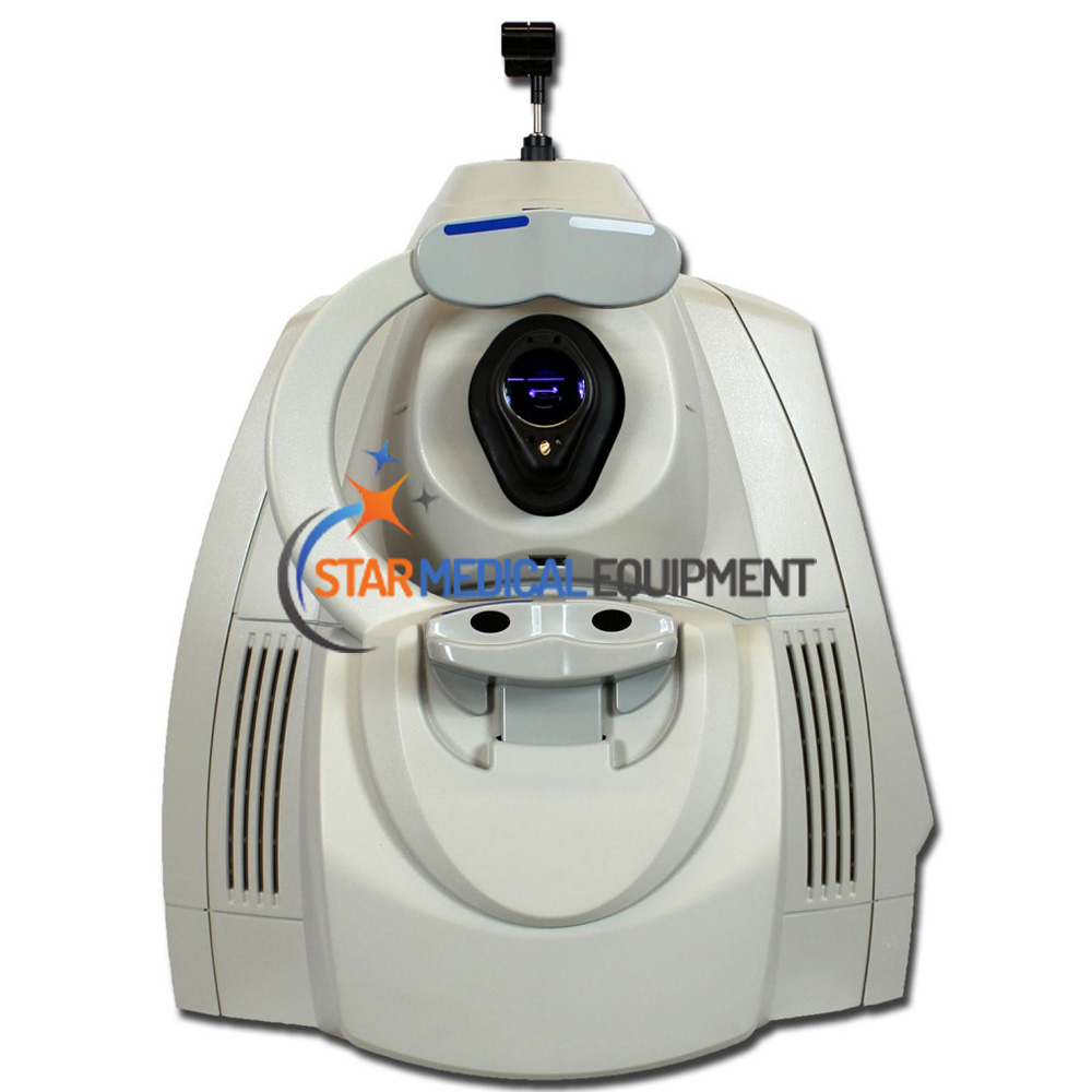
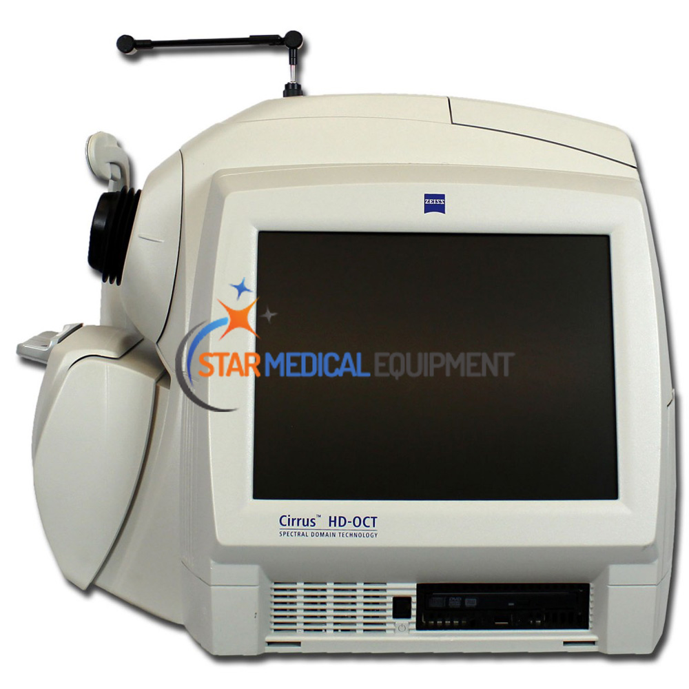
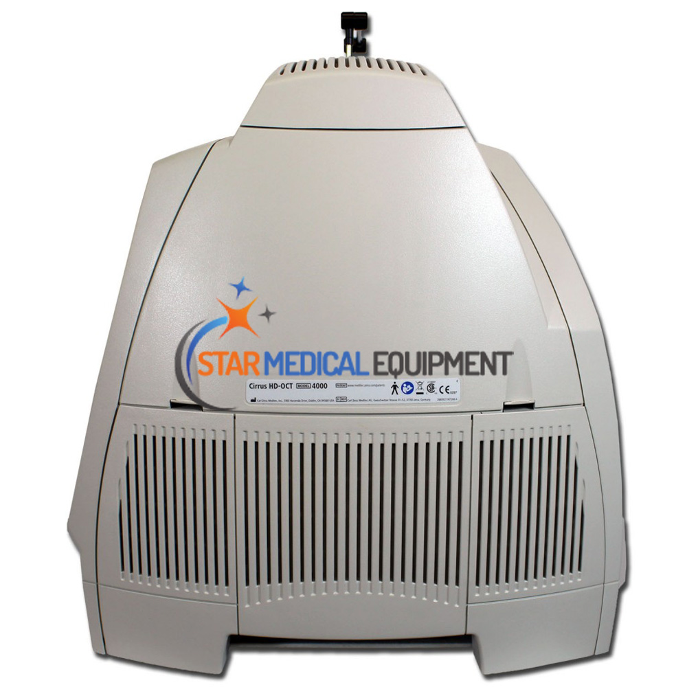
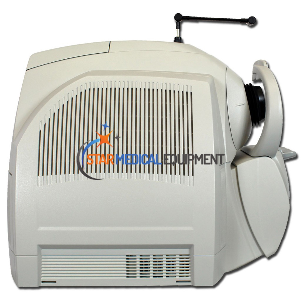
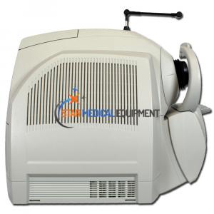
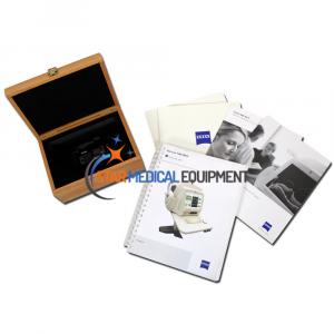
Zeiss Cirrus HD-OCT 4000 Corneal Topographer
Zeiss Cirrus HD-OCT 4000 Corneal Topographer
$9,000
Product Description
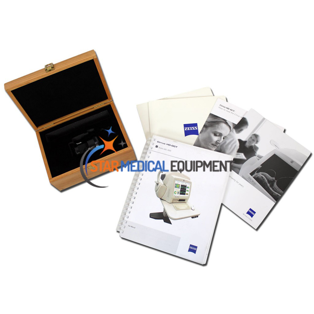
Sale Pre-owned Zeiss Cirrus HD-OCT 4000 Corneal Topographer enables examination of the posterior and anterior of the eye at an extremely fine spatial scale, without surgical biopsy or even any contact with the eye. Operatirng system Windows 7 installed (Upgrade to Windows 10 US$1,700).
Cirrus HD-OCT 4000 enables repeatable visualization of clinically relevant anatomy with exact correlation between the OCT scan and the fundus image.
Comprehensive navigational tools ensure efficient and simple operation.
Designed for efficiency
- Small footprint and integrated design are ideal for crowded or busy practice
- 90 degree orientation facilitates observation of patient throughout exam
- Advanced optics aid in the examination of patients with cataracts. Dilation is not required even for pupils as small as 2.5 mm
- Mouse Driven Alignment™ delivers superior image capture and analysis in just a few clicks, resulting in reduced chair time for the patient
- Auto Patient Recall™ assures patient position and instrument setting are repeated from previous visit
Cirrus HD-OCT 4000 Specification
| Scan Patterns | |
| Macular Cube 200 x 200 Combo | 200 horizontal scan lines comprised of 200 A-scans |
| Macular Cube 512 x 128 Combo | 128 horizontal scan lines comprised of 512 A-scans |
| 5 Line Raster | 4096 A-scans per B-Scan (adjustable length, spacing and orientation) |
| Fundus Imaging | |
| Scanning system | Line scanning ophthalmoscope |
| Field of view | 36 x 30 degrees |
| OCT Imaging | |
| Methodology | Spectral domain |
| Scan speed | 27,000 A-scans / sec |
| A-scan depth | 2.0 mm (in tissue) |
| Axial resolution | 5 μm (in tissue) |
| Transverse resolution | 15 μm (in tissue) |
| Internal Computer | |
| Operating System | Windows 7 |
| Processor | Intel® Pentium® Core 2 Quad |
| Memory | 4GB |
| Hard drive/internal storage | 750GB |
| Electrical | |
| Power | 100–120V~, 50/60Hz, 5A 220–240V~, 50/60Hz, 2.5A |
| Physical | |
| Size (L x W H) | 66.0 x 43.2 w 53.3 cm (26 x 17 x 21 in.) |
| Table footprint (L x W) | 99.1 x 55.9 cm (39 x 22 in.) |
| Software | |
|
|
